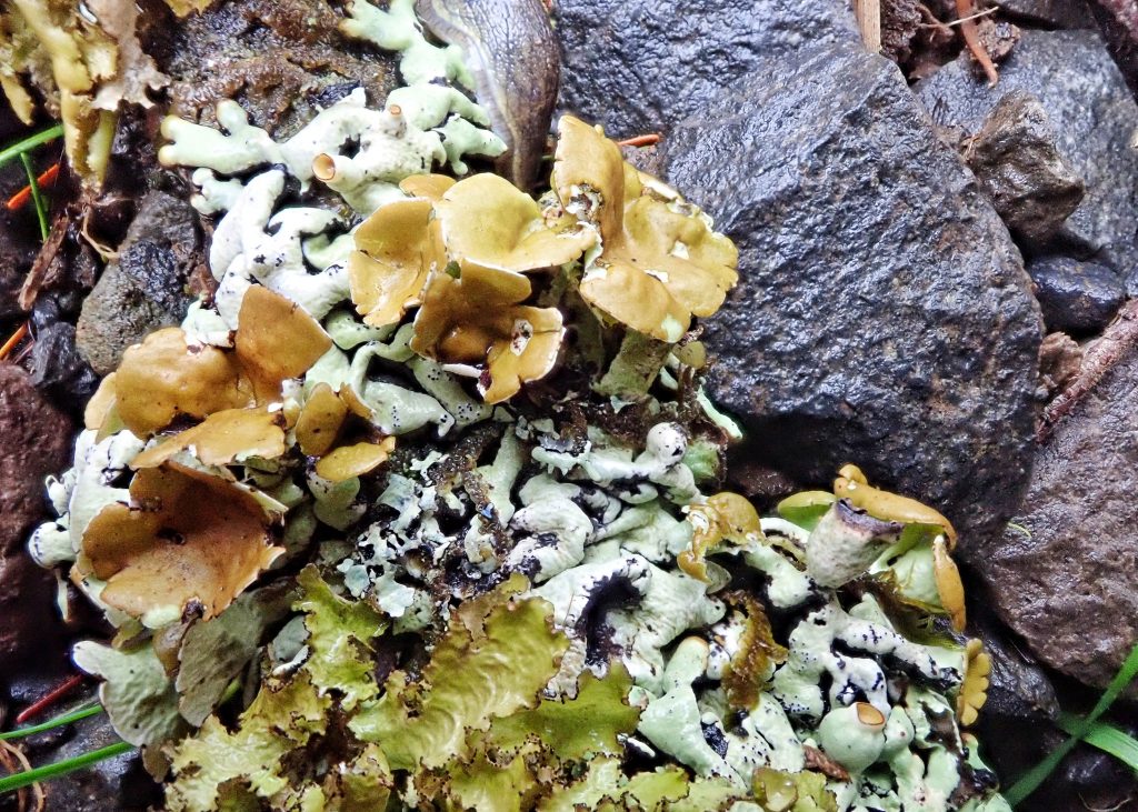
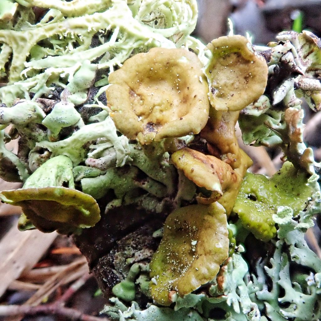
Finding this specimen on a windfall conifer branch sparked lots of confusion for this recreational naturalist! My first major error was that I became convinced that the apothecia were actually mushrooms. When I found it I did think they were apothecia, and as soon as I got home I looked at all of the photos I could find of the apothecia of Hypogymnia sp., but nothing matched. I even looked to see if they could be apothecia for other nearby species. But nothing clicked. My problem, besides lack of experience, was that I thought I knew that apothecia have corticular tissue, of the same color as the cortex, surrounding the cup, and that the cup doesn’t runneth over the sides.
So I posted it in a mushroom group, where Rand Workman had the unmitigated gall to give me the correct answer, that it was apothecia from the Hypogymnia. And then, from my wealth of inexperience, I proceeded to argue with them. And when Claire Whittaker chimed in, saying Rand was right, I argued with them also. Until they showed me a picture of older, waterlogged apothecia on a specimen of Hypogymnia, which looked very much like my specimens. I would guess that the bursting of my bubble made an audible ‘POP’. After confirmation from a few other sources I returned to the myco group and ate my raw, unseasoned crow. But I wasn’t done making mistakes.
That evening I went to visit my good friend Craig Sondergaard, since I have recently purchased some chemical reagents online, and he had agreed to split them with me. Neither of us had done spot tests of lichens before, but I had decided to make the leap because I’ve come to realize that if I want to be certain of my lichen identifications I am going to have to do spot tests. And Craig just likes to do cool scientific stuff. Spot tests are fairly straightforward, once you have the chemicals and the formulae, and involve putting a drop of the reagent on either the cortex or the medulla (that’s actually one of the hardest parts for me-exposing the medulla by tearing off a piece of the cortex, without completely mangling both), and waiting to see what, if any, reaction there is (click here for a great discussion of, and resource for, spot tests of lichens).
There are a couple other issues for me with spot testing. One of them is that these chemicals are hazardous, and a level of care that I don’t normally exercise is required to ensure that I don’t do anything stupid. The other is that I am colorblind. This is a real problem with weak reactions anywhere in the red spectrum, and was exacerbated by the fact that my buddy Craig is at least as color challenged as I am. Both of us struggle mightily with anything that is green or red, unless they are bright, solid, absolutely middle of the spectrum colors. When identification is heavily color dependent, it’s a real case of the blind leading the blind, which probably sounds a bit like a nerdy Abbott&Costello routine to the fly on the wall.
But things went fairly smoothly when spot testing my Hypogymnia. We both got a nice K+Y (K=potassium hydroxide, += positive, Y= yellow) reaction from the cortex, which helped confirm that it was Hypogymnia. Then we did a few more tests with separate chunks of the thallus, and got strong P+Red reactions from the medulla.
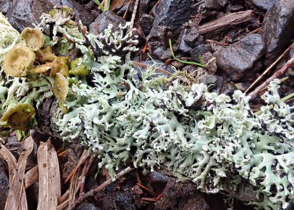
What wasn’t going smoothly was that I was finding masses of soredia, along with the aforementioned apothecia, and Hypogymnia is an either/or genus. Nothing really has both soredia and apothecia. I examined the piece I was working on, but it certainly did not pull apart, and I couldn’t see anything that made me think I had two organisms. And since the bursting lobe tips are diagnostic for Hypogymnia physodes (profiled yesterday), as was the P+R medulla, and it does, albeit rarely, have apothecia, I concluded that that was what I had, and moved on to the next lichen.
I was far from sure and fired off an email to Bruce McCune as soon as I got home. Who responded that, yes I had H. physodes, but I also had one of the fertile Hypogymnia. And, ironically, it turned out that part of the confusion was actually the solution, because Hypogymnia enteromorpha is also P+Red medulla, and is the only apothecate Hypogymnia with this growth form that is P+R! And rather than confirming H. physodes, as I’d thought the previous evening when I did a spot test on a chunk of thallus with apothecia growing from it, it now confirmed H. enteromorpha. So, mystery solved, ego humbled, and knowledge gained. That’s a good weekend!
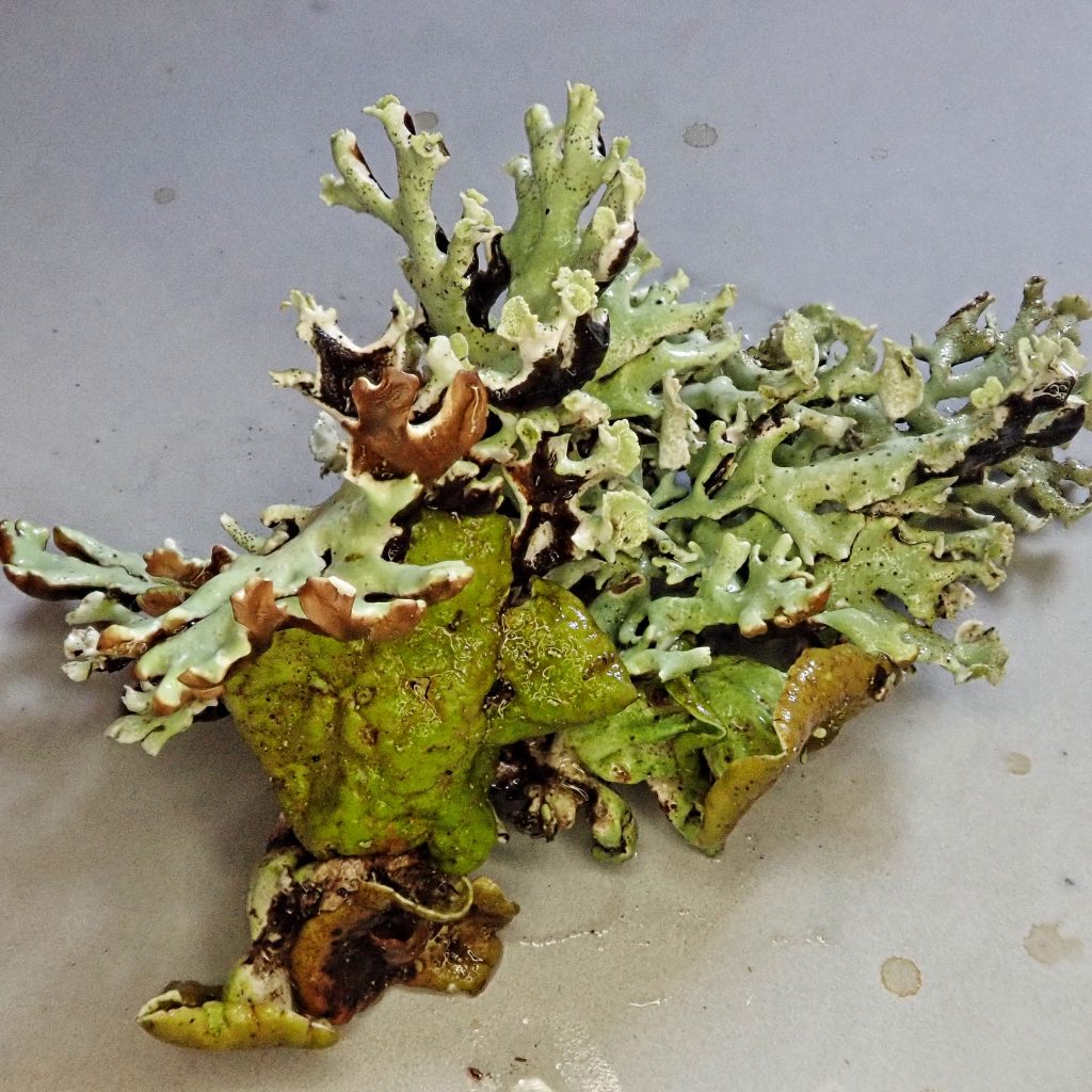
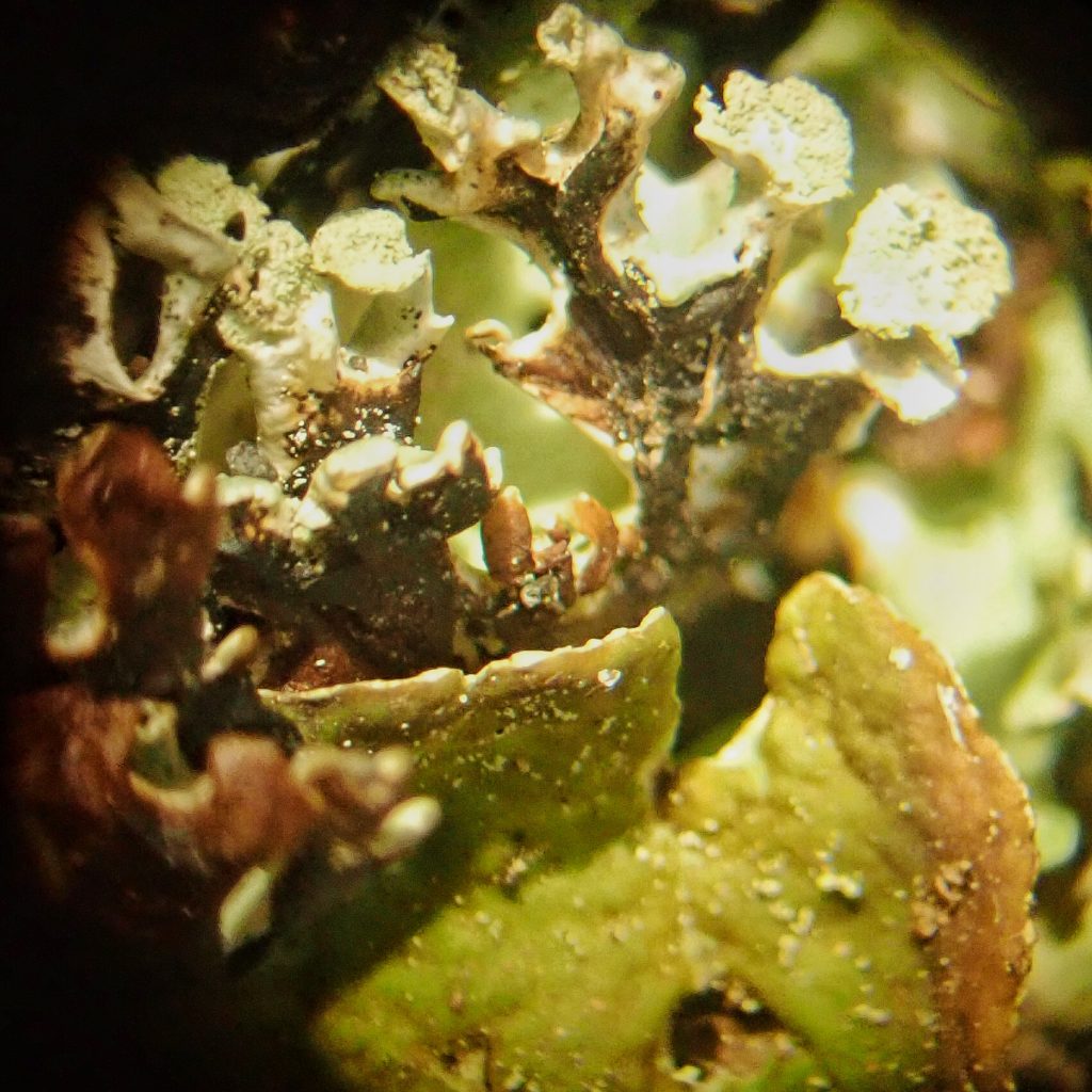
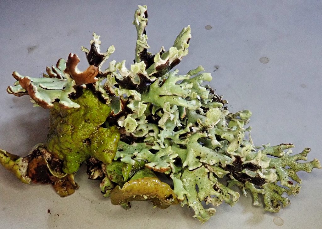
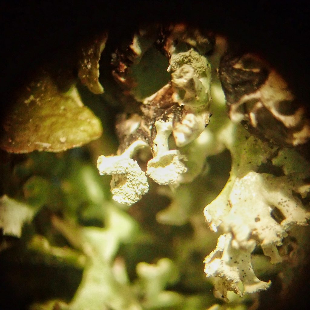
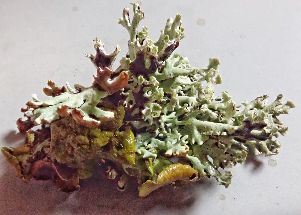
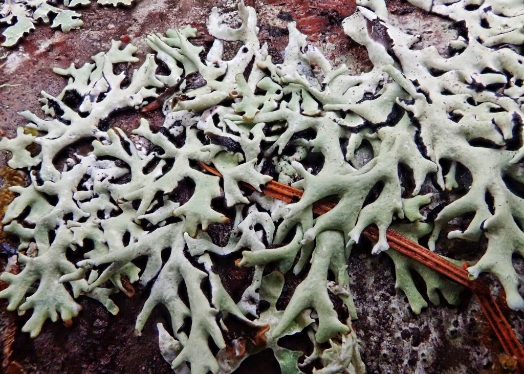
Description– Hypogymnia enteromorpha can be a fairly large lichen, although the specimen I found was not; greyish white to greenish grey on top, black on the ventral side; lobes 3-6mm wide, with constricted areas, and with bud-like, perpendicular side lobes, and lobules at the end; lacks soredia and isidia, but has brown/tan apothecia, and abundant pycnidia (tiny black, asexual spore producing structures); floor and ceiling of lobe interiors are dark; medulla P+R, KC+R (pinkish).
Similar species– H. apinnata is very similar and was long considered to be the same species (Goward/McCune; 1993). It usually has no, or just a few, bud like side lobes, and its medulla is P-, KC- ; H. occidentalis tends to have a wrinkled surface, and it’s medulla is P-.
Habitat-Found on bark and wood of conifers, hardwoods, and other woody substrates in areas with oceanic influence. Not found on rock or soil.
Range– Primarily in the Cascades and west to the Pacific; also in the Rockies.
Etymology of names–Hypo- means under or below in Greek, and -gymnia means naked, bare in Greek. This refers to the lack of rhizines in this genus. The specific epithet enteromorpha is also from the Greek and means ‘having the form of an intestine’, which probably references the hollow tubes of the lobes, and the way they are periodically constricted.
http://linnet.geog.ubc.ca/Atlas/Atlas.aspx?sciname=Hypogymnia%20enteromorpha
https://lichens.twinferntech.net/hyna/species/Hypogymnia_enteromorpha.shtml
https://lichens.twinferntech.net/pnw/species/Hypogymnia_enteromorpha.shtml
http://www.sharnoffphotos.com/lichensC/hypogymnia_enteromorpha.html
https://www.jstage.jst.go.jp/article/cpb1958/24/7/24_7_1596/_article
https://andrewsforest.oregonstate.edu/sites/default/files/lter/pubs/pdf/pub1517.pdf

So interesting that there is such a thing as chemical tests in the field in order to identify these things. Also amazing you can do all these identifications while red-green colorblind!
My wife is very helpful! And there are often other traits one can go on.
Hey Dan, to expose the medulla of a lichen, the easiest way is to get a pack of double edge shaving razor blades. Hold one between you fingers and snap it into two pieces giving you a single blade. With this you can cut away the cortex to expose the medulla. The blades break easily, but do be careful.
That’s basically what I do, Steve. The problem is that I have indelicate meathooks for hands, and I’m clumsy as an ox. But I muddle through😀
“Wealth of inexperience” 🤣👍